
Wednesday, 07 January, 2026г.


Где искать: по сайтам Запорожской области, статьи, видео ролики
пример: покупка автомобиля в Запорожье
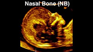
Nasal bone assessment and absent nasal bone at 11-13 weeks
 У вашего броузера проблема в совместимости с HTML5
This is a training ultrasound video about assessment of the fetal nasal bone (NB) as part of the Down's syndrome screening at 11-13 weeks' gestation.
Protocol for assessment of the fetal nasal bone:
The gestation should be 11-13+6 weeks and the CRL 45-84 mm.
The magnification of the image should be such that the head and upper thorax occupy the whole screen.
A mid-sagittal view of the fetal profile should be obtained.
The ultrasound transducer should be parallel to the direction of the nose and the probe must be gently tilted from one side to the other of the fetal nose.
When these criteria are satisfied, three distinct lines should be seen at the level of the fetal nose:
The top line represents the skin.
The bottom one, which is thicker and more echogenic than the overlying skin, represents the nasal bone.
A third line in front of the bone and at a higher level than the skin represents the tip of the nose.
The nasal bone is considered to be present if it is more echogenic than the overlying skin and absent if it is either not visible or its echogenicity is the same or less than that of the skin.
The same mid-sagittal view of the fetal profile at 11-13 weeks of pregnancy is also used for nuchal translucency (NT) thickness measurements and intracranial translucency (IT) assessment.
At 11-13+6 weeks the nasal bone is considered to be absent in about:
Euploid fetuses 1-3%
Fetuses with trisomy 21 60%
Fetuses with trisomy 18 50%
Fetuses with trisomy 13 40%
Absence of the nasal bone is more common if:
The gestation is 11 than 13 weeks
The fetal nuchal translucency is high
The mother is Black
Теги:
fetal nasal bone nasal bone absent nasal bone scan nuchal translucency intracranial translucency Down syndrome trisomy 21 trisomy 18 trisomy 13 how to do ultrasound technique Down's syndrome testing Downs syndrome screening nasal bone ultrasound yt:stretch=16:9
Похожие видео


Мой аккаунт