
Tuesday, 13 January, 2026г.


Где искать: по сайтам Запорожской области, статьи, видео ролики
пример: покупка автомобиля в Запорожье
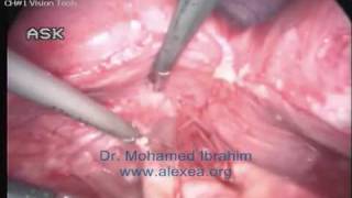
14 years old patient case presentation
 У вашего броузера проблема в совместимости с HTML5
14 years-old female presented to the Clinic, complaining of severe pelvic pain at one side for one month; the pain started abdominally since 2 years.
The patient stated that she didn't menstruate yet.
Computed Tomography revealed: Marked distention of the Vagina, Bicornuate uterine cavity and multiple pelvic and abdominal multilocular peritoneal collections.
Diagnostic laparoscopy was done and revealed the following:
- Bicornuate uterine cavity.
- Huge bilateral Heamtosalpinyx.
- Both ovaries look normal.
- Few clear free fluid collection.
- Appendix looks free.
- Aspiration showed choclate material.
Following Investigations were done:
- Carcinoembryonic antigen (CEA 125) is 450.
- Hormonal profile:
LH: 6.1
FSH : 2.5
Estrogen: 137
Surgical procedure was done later;
Introduction of Hysteroscope (2.9 mm) through the hymenal opening was done, the vaginal part of the cervix was seen, trials to pass through the cervix but no continuation could be achieved.
Laparoscopy was done;
- Coagulation and incision of Left Tube.
- Suctioning and evacuation of the retained blood.
- Coagulation and incision of Right Tube.
- Suctioning and evacuation of the retained blood.
- Complete adhesiolysis and Mobilization of Uterus, Rectum and both adenexea : Now uterus, adenexea and the Douglas pouch are completely free.
- Uterus is bi-cornuate, the right side looks more in line and continous with cervix.
The patient was advised to:
1) Progesterone pills: to stop period
2) Second look after 2-3 months
3) Hysteroscopy to differentiate between bicornuate uterus or non communicating horn
Pathology results for the Left Adenexal Mass showed no signs of Malignancy.
Теги:
14 years 14 years old case gynecological case gynecology case presentation case presentation alexea alexandria alex laparoscopy endoscopy gynecology gynecological MOhamed Ibrahim Dr. Mohamed Ibrahim Mohamed Ibrahim
Похожие видео


Мой аккаунт